There have been few changes in colposcopy in recent decades despite many advancements in other areas of cervical cancer treatment. HPV testing and vaccination have offered a new dimension to cervical cancer screening and prevention but few adaptations have been made to the practice of colposcopy.
Visual imaging of the cervix remains an essential part of any cervical cancer diagnosis. If a Pap Smear or HPV test returns a positive result, it is still necessary to visualize the cervix to assess lesions and take biopsies.
The recent development of the Automated Visual Evaluation (AVE) algorithm promises to screen for cervical cancer using a single image of the cervix, returning a result at the point of care, removing the need for the delays, expense, and loss to follow up issues caused by current primary screening methods. However, even with this significant breakthrough in screening methods, visualizing the cervix will still be necessary to apply the algorithm. (Once, the AVE algorithm has classified an image, additional tests for a specific diagnosis, such as CIN 2 vs CIN 3, and for the management of advanced disease will still be needed.
MobileODT set out to explore new methods of cervical visualization to assess whether improvements could be made to existing methodologies.

What is multispectral imaging?
Multispectral imaging captures data at specific wavelengths across the electromagnetic spectrum. It enables the collection of additional information the human eye fails to capture by measuring light in a small number of spectral bands (colors). It was originally developed for space-based imaging.
Multispectral imaging has found applications in an ever-growing range of industries from the military (searching for landmines), mineral exploration, archeology, art restoration, as well as biomedicine.
Multispectral imaging in the medical field
Multispectral imaging has made a significant impact in medical diagnostics. Imaging is an essential part of the medical field, especially with advances in MR- and CT- based imaging methods. However, these techniques only show anatomical changes, not molecular variations. As many diseases begin with molecular alterations, a method that easily demonstrates such changes would offer a significant improvement in early diagnosis.
One early medical application of multispectral imaging is measuring oximetry (blood content and oxygen saturation) in superficial tissues. Spectral imaging is used to assess oximetry without requiring contact with the patient or any injectable contrast agents. (Oximetry is measured by analyzing the spectral composition of light diffusion reflected.) Instead, it uses visible light and a spectrometer to image and quantify light absorption of hemoglobin molecules. This benefits patients with compromised vascular systems such as those suffering from diabetes, peripheral vascular disease and limb ischemia. In extreme cases, correct detection of oximetry can enable preservation of limbs.
Multispectral imaging for cervical cancer
Studies looking into the possibility of using multispectral imaging for the detection of cervical cancer show great promise. The potential to detect cancer based on viewing molecular changes instead of relying on the opinion of a clinician, as is the case in Visual Inspection with Acetic Acid (VIA) procedures, or waiting for the results of an invasive biopsy, has incredible advantages.
For any such diagnostic imaging to reach a practical application, a cost-effective solution that can be integrated into a colposcope is required.
MobileODT’s EVA System is a digital colposcope that incorporates both medical-grade imaging and a robust software application that allows for secure documentation and image storage. If multispectral imaging was incorporated into the portable EVA System then it could become a viable option in a wide range of healthcare scenarios.
Developing practical applications for multispectral imaging
In order to develop a practical application for cervical cancer leveraging multi-spectral data analyses based on light transport theory, MobileODT has been conducting parallel studies using both external skin imaging and imaging of cervical tissues.
Our work exploring the potential to create a low-cost system that could assess tissue composition based on light scattering and absorption. This, in turn, would enable the detection of cancerous molecules in the tissue. At this preliminary stage of development, we assessed erythema on external tissue (the forearm) as a proof of concept.
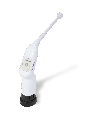
We purchased common components used in consumer electronics including a tablet and an area-scan camera. Other components were combined to create a 13-LED illumination board. Digital images were recorded for each wavelength. Every multispectral cervical data set was uploaded to the cloud for analysis.
In total the cost of parts was under US$1,000, a production cost two times less expensive than anything commercially available.
Once we had built a functional low-cost multispectral imaging system, we proceeded to test our hypothesis that healthy tissue would display less scattering as light is absorbed into the microvasculature. Our findings were consistent with the hypothesis.
MobileODT’s preliminary work into multispectral imaging of cervical tissue was presented in a talk at the Optics and Biophotonics in Low Resource Settings conference at SPIE’s Photonics West 2018 conference. In this study, patients who were undergoing colposcopy or LEEP were recruited to participate. Once acetic acid had been applied, the cervix was imaged with the multispectral device and then the procedure continued as standard. The data was processed offline and the site of any biopsies was recorded.
Each multispectral data set was captured in under five seconds, calculations of blood content, oxygen saturation, and scattering were made. As biopsy sites were tracked, it was possible to compare the output of the spectral data with histopathology. We found that the estimated values of blood content, oxygen saturation, and scattering were within the range we expected for epithelial tissues.
See the poster for more details and data>>
The future of multispectral imaging in cervical cancer
As we demonstrated in our study, it is possible to create an affordable, effective multispectral imaging system for use in the examination of cervical tissues. MobileODT’s work is ongoing as we explore the many possibilities that it opens up. When developed further this system has the potential to significantly impact the diagnosis of many topical forms of cancer, as well as detect other conditions of the cervix and lower genital tract, such as preterm birth, fertility, and other pregnancy-related issues.
MobileODT is committed to exploring new adaptations of technology to bringing affordable healthcare solutions to clinicians around the world. Multispectral imaging is just one of the many areas we are pursuing. Learn more about our work with augmented intelligence to aid in cervical cancer detection>>