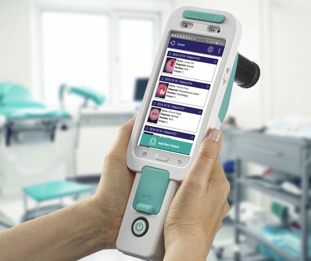
A new research study may help put effective cervical cancer treatment within the reach of millions of women in low resource settings globally. Published in the British Medical Journal, the prospective study compared methods of cervical cancer screening with the aim of finding the optimal method for low resource settings, in terms of cost and efficacy.
The study was led by Dr. Andrew Goldstein, Obstetrics and Gynecology, The George Washington University School of Medicine and Health Sciences, and The Center for Vulvovaginal Disorders, Washington DC; and Dr. Sovannara Thay, Department of Gynecology, Sihanouk Hospital Center of Hope, Phnom Penh. It was made possible through the support of the Gynecologic Cancer Research Foundation.
The cervical cancer burden in low resource settings
Despite advances in cervical cancer prevention and detection with HPV vaccination and testing, an estimated 311,000 women globally die from the disease every year. 84% of these deaths occur in low- and middle-income countries. Early detection is the key to reducing mortality.

There are many challenges facing the implementation of effective early detection programs in low resource settings, including the human and physical capital to implement traditional cytology and HPV based programs that require laboratories and expert technicians to interpret results. Besides the need for the most cost-effective solution possible, any program must also consider the societal realities for women in these communities. There is a significant burden placed upon women accessing healthcare services.
Even when free services and screening camps are provided, the time taken to attend them frequently creates a number of challenges including loss of income, travel expenses, childcare challenges and loss of productivity. Thus an optimal method is one that can be implemented with limited resources and meets the needs of women to actually uptake the service.
Choosing a suitable research environment – Cambodia
In order to comprehensively test the efficiency of cervical cancer screening methods, the study required an environment that allowed for access to a high level of cervical cancer and precancer incidence.
Cambodia has one of the world’s highest cervical cancer rates. The pervasive sex trade, with up to 80% of Cambodian men taking part, results in rampant HPV infection both among women directly participating in the trade and family members of men who do so.

Two hundred and fifty women were recruited to take part in a study from the National Center for HIV/AIDS Dermatology and sexually transmitted disease (STD) cohort, the Sihanouk Hospital Center of Hope’s Rural Outreach Teams and the Pochentong Medical Center. In order to compare methods in both standard and high-risk populations, the sample was divided equally among HIV positive and negative populations.
How cervical cancer screening methods were evaluated
Both specificity and sensitivity were evaluated. Cathy Sebag, Product Manager at MobileODT, who helped provide technical support for the research study, explains, “Women in a low resource setting are unlikely to have access to regular screening options, therefore it was essential that no woman is ‘sent home’ with a false negative result. In a high resource setting, a false negative result will likely be picked up in the next round of screening.
For women in low resource settings the next screening might never happen.”
A more accurate screening method, that differentiates between high and low-grade lesions also allows for the most suitable treatment method to be selected. “We obviously want partners to perform the more invasive procedures only when necessary, such as LEEP and even surgical intervention for advanced cases. Although low resource programs often go on the assumption that over-treatment is better than under-treatment.
Clearly, best is the correct treatment to achieve maximum health outcomes with minimum harm to the patient by creating the screening method that allows more precise diagnosis without increasing cost or expertise needed,” Sebag says.
Methods compared
Four screening methods were compared in this study:
HPV self-sampling
The ability for women to perform an HPV self-sample test has been lauded as a significant step forward in cervical cancer screening. Self-sampling removes much of the potential embarrassment associated with sampling by a clinician, and the number of providers who need to be trained and available to take samples from speculum exams. It is cost effective as it requires no clinician participation beyond educating the women on how to perform the self-sample. Positive HPV results would be followed by traditional colposcopy and biopsy, or in some cases VIA, before moving on to any necessary treatment.
HPV sampling by a clinician
The standard of care clinician-collected HPV sample through a speculum exam was compared to the overall accuracy of self-collected specimens. Previous studies have shown that healthcare provider-collected cervical samples have significantly higher accuracy for detecting high-grade CIN as compared with traditional pap smears. While self-collected samples require much fewer experts to reach many more women, different geographic locations need to be investigated to ensure this is an acceptable method, and that women appropriately get the sample from the cervix needed for an accurate result, compared to that expert test. Again women showing positive HPV results would then have traditional colposcopy and biopsy, or VIA, before moving on to any necessary treatment.
VIA
Visual Assessment with Acetic acid is the standard cervical cancer screening in many low resource settings. It allows for immediate treatment of any lesions found but as it relies on the naked eye assessment of an experienced clinician it is potentially problematic. However, the immediacy of the treatment may allow for an increase in positive health outcomes as the loss-to-follow-up challenge is almost entirely removed in a VIA screen and treat scenario.

Digital Colposcopy (using the EVA System)
The advent of portable digital colposcopes has been enthusiastically welcomed in cervical cancer screening programs in low resource settings. It allows for enhanced visualization of the cervix and can be followed by immediate treatment as with VIA. The main difference is that the magnification allows the provider to distinguish beyond normal and abnormal, and to distinguish between low and high grade to decide the most appropriate treatment This potentially increases the accuracy of VIA while still removing the loss to follow up issues that might be found with HPV testing.
Results
There were some surprising results found in the study. Self-sampling by patients was found to have a greater level of accuracy than by non-expert clinician. “We think that women were worried not to perform the self-sampling correctly, so were vigorous in their sample collection,” Cathy Sebag surmised. In fact, the study showed 95.2% of Cambodian women are comfortable obtaining these specimens.
The VIA track found that it was very difficult to distinguish between a high or low-grade lesion with the naked eye.
In some cervical cancers screening clinics in low resource settings, this would likely result in over or under treatment of the lesion. In the case of overtreatment, this might leave women needlessly suffering the side effects of LEEP. In under treatment, not all of the problem areas may have been addressed or removed, and cancer may develop if the patient does not return for regular screening, which is common in this setting.

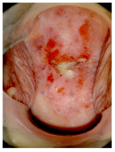
Figure 2. Image captured with the EVA System.
While digital colposcopy is also visualization, it had significant advantages over VIA alone. While previous studies regarding VIA have shown that physicians are more likely to detect CIN2+ than nurses, DC has several benefits over VIA that can help reduce the difference between nurses and physicians. Second, the DC images can be used for continued education of nurse/midwives and for quality control.
A 100% correlation was found between the physician’s impression of the type of lesion and later biopsy analysis. This has the potential to significantly reduce costs to screening programs.
Condensing the entire process into one patient visit dramatically reduced the burden of accessing healthcare for the women being screened, giving women a treatment modality that suits their needs.
Further studies
This study opens the way for cervical cancer programs in low resource settings to confidently deploy digital cervicography with the EVA System as their treatment model.
The results of this study might suggest potential modifications of the current cervical screening strategy that is currently being employed in Cambodia on a limited basis:
A large number of Cambodian women could collect self-collect swabs; the careHPV system, which is highly portable, could be used to rapidly test these specimens. In addition, the cost is relatively inexpensive at only approximately US$4 per specimen.
1. The second component of this proposed strategy would be to perform DC on hrHPV+ women with the EVA system. Given the shortage of trained gynecologists in Cambodia, DC can be performed by trained nurses or midwives.
2. A potential third component of a rapid, and cost-effective, screen-and-treat strategy might be to use the clinical impression derived from the EVA System images to determine immediate treatment. Specifically, women with suspected CIN1 lesions would be immediately treated with cryotherapy and those with suspected CIN2+ lesions would have LEEP
3. The strategy is unique from WHO guidelines, which recommends cryotherapy in all abnormal cases, to make sure the patient gets the appropriate treatment based on the higher quality exam results.


