 By Molly Webster and Vikram Sheel Kumar
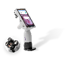
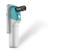
The Pap smear has been used to test women for cervical cancer since its invention in 1928. With a simple swipe and smear, doctors have been able to use microscopic observation to detect cancerous activity. Because cervical cancer is one of the most preventable forms of cancer, the Pap smear revolutionized how we deal with the disease. Yet, worldwide, 275 000 women (1) still die from cervical cancer each year. That’s because although the Pap is an efficient test, it requires elements such as facilities, follow-up visits, and trained cytology technicians, making it difficult to use in low- to middle-income neighborhoods. In these areas, some of which are right here in the US, visual inspection with acetic acid (VIA)3 has become the norm in cervical cancer screening. In this procedure, the cervix is washed with acetic acid, and a physician scans the cervix with his or her own eyes, without any magnification, looking for white spots, which might possibly be a clump of cancerous cells. The problem with VIA, however, is that it can offer an often imprecise and delayed picture. If a physician can actually see the cancer on the surface of the cervix, that would indicate the tumor has become large enough to burst through the skin’s surface, from the deeper tissues below, meaning it is very advanced. And, white doesn’t always mean cancer. So, although VIA has reduced cervical cancer mortality rates by some 30% (2), it has a false-positive rate that can hover near 83%. What would be ideal, says David Levitz, cofounder of MobileOCT, a technology company, is a different type of screening test that can enhance a physician’s vision beyond 20/20. MobileOCT is working on just such an optical cervical cancer screening technology, optical coherence tomography (OCT). [More here]


