By using visual assessment tools to educate patients at the point of care, clinicians can improve patient outcomes. Studies indicate that showing patients real-time imaging and 3-D models to teach patients not only improves medical literacy but also reduces fears, improves quality of life, increases follow up and retention, and builds greater trust in physicians.

3D examples reduce patient fears and improve quality of life
A 2013 study observed the effects of educating patients about their diagnosis using 3D models in relieving patient concerns related to misunderstandings of the severity of the diagnosis. The participants included twenty women with pelvic floor disorders (UI or POP) between the ages 31-87.
Interviews conducted before and after the initial consultations with physicians revealed that education through the use of models has a significant impact on patient perceptions and understanding of their condition. Prior to treatment, patients lacked an understanding of both their diagnosis and treatment options. During interviews, women expressed fears regarding the severity of the diagnosis and lack of control over their condition.
A practical education session during their consultation led most participants to gain a much better understanding of their treatment plan. Physicians used pelvic models to teach patients about their anatomy and treatments plans. As a result, patients expressed relief from their initial concerns, feeling more informed and in control. The outcome shows that informing patients reduces worry and leads to “less anxiety, less depression and better quality of life”.

Visual assessment tools enable patients to better understand their bodies
Another study looked into patient attitudes in seeing their own X-ray and CT scans, revealing that patients who see images of their bodies gain a stronger understanding of the diagnosis. They also receive validation of their sensory and emotional response to an illness or injury. In addition, showing images builds a stronger trust between the patient and clinician, increasing the likelihood for the patient to follow up for additional treatment.
Meanwhile, not showing images can have adverse effects, and patients take notice. When physicians do not take it upon themselves to share images, patients may take it upon themselves to do their own research. Patients “read into’ it if doctors do not share any images. Conducting their own research could lead to misinformation and increase patient fears and confusion on health conditions and treatment.
Guided procedures improve patient experience and increase follow up
One study looked at patients who observed themselves while receiving ultrasound-guided injections. In this case, 92% of the participants felt that observing the ultrasound images in real-time during a guided procedure made them feel more at ease during the injection. Also, 95% of patients were very likely or somewhat likely to undergo repeat treatment, if needed. Patients also noted that they found this experience better than the unguided procedure.
Showing medical images during procedures improves patient trust in clinicians
Presenting medical images during exams and procedures also improves patient trust in clinicians. An experimental study compared the impact of viewing no image, a 2D medical image, and a 3D medical image during diagnosis.
The results concluded that patients preferred the 3D images the most alongside diagnosis. Further analysis also reveals that medical images aid patient understanding, recall, and trust in medical information.
EVA WELL – subtitles from Peter Gorne on Vimeo.
Empowering women to better understand reproductive health
Being unable to see what is happening during a gynecological exam, can lead some women to have fears and misunderstandings of health conditions, diagnosis, and treatment plans.
Patients experience improved outcomes by seeing real-time imaging and 3D models during gynecologic exams. To encourage patient education, different organizations work alongside clinicians to provide visual tools to help inform patients at the point of care.
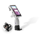
MobileODT’s EVA System provides another way for patients to learn about their bodies by viewing real-time images of their anatomy at point of care. The visualization tool allows clinicians to show images and videos during an exam, reassuring and educating patients about any findings and progress in treatment. The level of engagement provided by the EVA System encourages patients to take an active part in their care, which increases retention and follow-up.
Outside of the clinic, the Beautiful Cervix Project is a non-profit that offers an at-home service for testing STDs, vaginal imbalances, and HPV. In addition, the website offers pictures inside the vagina as an educational tool for women.
By showing images and using visual assessment tools, physicians can better educate women at the point of care. Knowledge of their own bodies and health conditions enables patients to understand their treatment plan (which will lead to more compliance) and increases patient trust and relationships with clinicians.
Read more:
Vagina or vulva- the importance of patient education